


Osgood-Schlatter disease
Definition
During your child's adolescent growth spurt, his or her bones grow rapidly. If your child is involved in a lot of running and jumping activities during this time, he or she is at risk of developing Osgood-Schlatter disease, an overuse syndrome that causes pain, swelling and tenderness over the bony prominence of the upper shinbone (tibial tuberosity) just below the kneecap. The condition is also referred to as tibial tuberosity apophysitis.
Osgood-Schlatter disease occurs more often in athletic kids than in nonathletes, affecting as many as one in five adolescent athletes. The condition commonly occurs in boys ages 13 to 14 and girls ages 11 to 12. Osgood-Schlatter disease is more common in boys.
Having Osgood-Schlatter disease can be frustrating, because your child may need to limit his or her running and jumping activity level for a short time. But Osgood-Schlatter disease is temporary, and as your child's bones finish growing, the pain should go away.
Symptoms
Signs and symptoms of Osgood-Schlatter disease include:
* Pain, swelling and tenderness at the bony prominence (tibial tuberosity) on the upper shinbone (tibia), just below the kneecap
* Knee pain that worsens with activity, especially running and jumping, and improves with rest
* Tightness of the surrounding muscles, especially the thigh muscles (quadriceps)
The pain varies from person to person. Some have only mild pain while performing certain activities, especially running and jumping. For others, the pain is nearly constant and debilitating. Osgood-Schlatter disease usually occurs in just one knee, but sometimes it develops in both knees. The discomfort can last from weeks to months and may recur until your child has stopped growing.
Causes
New bone forms from a cartilage growth plate (epiphysis) located at either end of the bone. Cartilage isn't as strong as bone, and stress on the growth plate can cause it to become swollen and painful, especially if your child is very active during his or her growth spurt.
Osgood-Schlatter disease is caused by activities that place repeated stress on the top of the tibia, the big bone in the lower leg, where the tendon of the kneecap inserts. During activities that involve a lot of running, jumping and bending — such as football, soccer, basketball, volleyball, gymnastics and ballet — the pull of the quadriceps can place tension on the band of tissue that connects the knee to the tibia (patellar tendon).
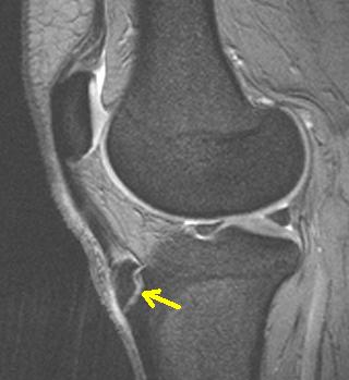
The patellar tendon may begin to pull away from the raised area on the tibia where it attaches (tibial tuberosity), resulting in pain and swelling. In severe cases, the tendon stretches to the point where it actually detaches from the tibia, and it may take a bone fragment with it.
----
and from wiki:
Osgood-Schlatter disease
Osgood-Schlatter disease or syndrome (also known as tibial tubercle apophyseal traction injury) is an inflammation of the growth plate at the tibial tuberosity, and is one of a group of conditions collectively called osteochondroses. The condition is named after the American surgeon Robert Bayley Osgood (1873–1956) and the Swiss surgeon Carl Schlatter (1864–1934), who independently described the disease in 1***.[1][2][3]
The condition occurs in active boys and girls aged 11-15[4], coinciding with periods of growth spurts. It occurs more frequently in boys than in girls, with reports of a male-to-female ratio ranging from 3:1 to as high as 7:1. It has been suggested the difference is related to a greater participation by boys in sports and risk activities than by girls.
The condition is usually self-limiting and is caused by stress on the patellar tendon that attaches the quadriceps muscle at the front of the thigh to the tibial tuberosity. Following an adolescent growth spurt, repeated stress from contraction of the quadriceps is transmitted through the patellar tendon to the immature tibial tuberosity. This can cause multiple subacute avulsion fractures along with inflammation of the tendon, leading to excess bone growth in the tuberosity and producing a visible lump.
The syndrome may develop without trauma or other apparent cause. But some studies report up to 50% of patients give a history of precipitating trauma.
In a retrospective study of adolescents, young athletes actively participating in sports showed a frequency of 21% reporting the syndrome compared with only 4.5% of age-matched nonathletic controls.[5]
Sinding–Larsen–Johansson Syndrome is an analogous condition involving the patellar tendon and the lower margin of the patella bone, instead of the upper margin of the tibia.
Symptoms
- Knee pain is usually the presenting symptom that occurs during activities such as running, jumping, squatting, and ascending or descending stairs. The pain can be reproduced by extending the knee against resistance, stressing the quadriceps, or squatting with the knee in full flexion. Pain is mild and intermittent initially. In acute phase the pain is severe and continuous in nature.
- Bilateral symptoms are observed in 20–30% of patients.
- The symptoms usually resolve with treatment but may recur as a new episode until skeletal maturity, when the tibial epiphysis fuses. Symptoms, however, may continue to wax and wane for 12–24 months before complete resolution. In approximately 10% of patients the symptoms continue unabated into adulthood, despite all conservative measures.[6]
Treatment
Diagnosis is made clinically,[7] and treatment is conservative with rest and simple pain reduction measures of ice packs and if required paracetamol (acetaminophen) or ibuprofen. The condition usually resolves in a few months, with a study of young athletes revealing a requirement of complete training cessation for 3 months (on average) and gradual resumption of full training by 7 months.[5]
Bracing or use of plaster of paris to enforce joint immobilization is rarely required and does not necessarily give quicker resolution.[8] Surgical excision may rarely be required in skeletally mature patients.[6]
After symptoms have resolved, a gradual progression to the desired activity level may begin. In addition, predisposing factors should be evaluated and addressed. Commonly quadriceps and/or hamstring tightness is present and should be addressed with stretching exercises. Training factors such as intensity and repetition should also be evaluated and addressed.


 Reply With Quote


 I go back to see the doc in another couple weeks. Anything you think I should look into/research meanwhile? Appreciate the insight
